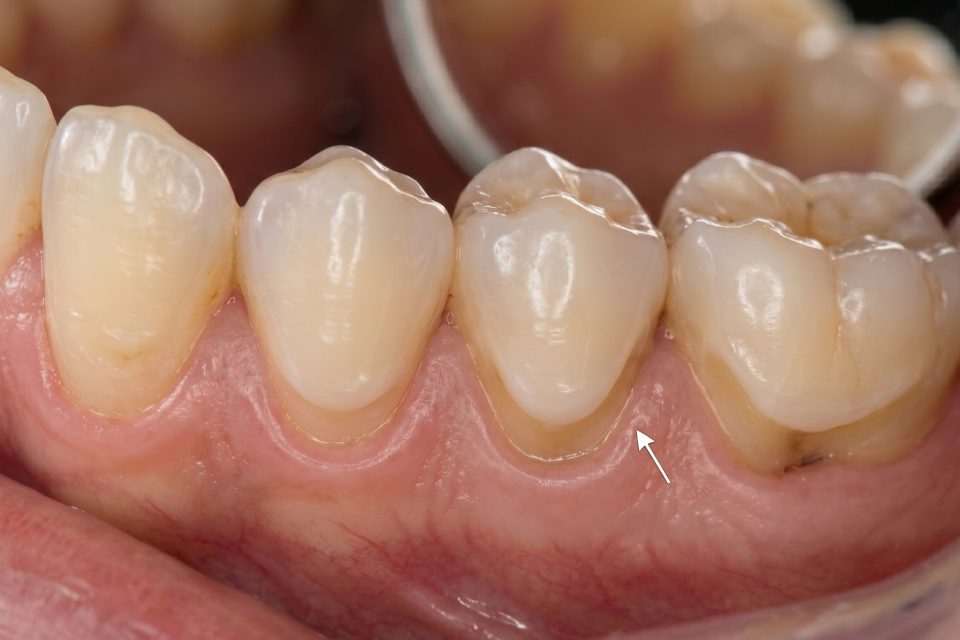
The term ‘abfraction’ or ‘abfraction lesions’ is not commonly known as it has only been introduced to dentistry in the early 90’s. Prior to that the condition, which has been around for a long time, was referred to as abrasion or erosion. Abfraction is a non-cavity related loss of tooth structure in the area where the tooth and gum join together. Left untreated, it can cause everything from fractured teeth and reduced enamel to total tooth loss.
What is Abfraction?
Abfraction is a non-carious tooth lesion and loss of tooth structure caused by mechanical stress like grinding and chewing. This repeated stress causes microscopic bending of the tooth which eventually leads to enamel and dentin breakdown at the cervical area. Abfraction presents as small V-shaped notches near the gumline and can look like dents in the teeth along the gumline. This pathological loss of tooth substance primarily affects the enamel and dentin. Unlike cavities, these types of lesions are usually smooth, hard and sharply defined. Although often confused with abrasion, which is tooth wear from brushing, or erosion (tooth wear from acids), abfraction is only driven by occlusal forces like bruxism or misaligned bite.
Abfraction Symptoms
Abfraction symptoms usually begin as quite painless but can start to become uncomfortable in time. Most people do not notice it initially but may become aware of these symptoms as the condition progresses:
- Sensitivity: The enamel loss at the gumline can cause tooth sensitivity from abfraction. The exposure of the sensitive inner dentin causes tenderness to hot, cold or sweet things. Dentin has tiny tubules leading to the nerve which causes sharp pain and sensitivity when exposed.
- Tooth grooves: Abfraction causes web shaped tooth grooves and notches near the gumline. Usually the first sign of visible presence of the grooves is small but can be seen as a slight yellow or brownish discolouration of the tooth near the gum.
- Pain when brushing: If you feel pain when brushing or touching a tooth, it may be a sign of abfraction. The teeth becoming sensitive to the touch is a sign of enamel loss and dentin deterioration.
- Food trapping: Food trapping is a common symptom of dental abfraction. The indentations caused by the abfraction create spaces where food particles can become trapped and accumulate which increases the risk of tooth decay and gum disease.
- Tooth fractures: Abfraction can lead to the symptom of tooth fractures if left untreated for extended periods of time. Because abfraction causes structural weakening of the tooth it is susceptible to fracturing. Tooth fractures may look like small hairline cracks or even a split tooth and are usually a sign that abfraction has advanced.
- Decay: Abfraction is a significant risk factor for developing tooth decay. In this case, tooth decay is not a direct symptom of abfraction but a potential secondary complication.
- Tooth loss: In the most severe cases, tooth loss can be a symptom of abfraction. Without intervention, the damage to the tooth structure becomes so severe that it destabilises teeth causing them to fall out. Should tooth loss occur, abfraction has advanced significantly.
Causes of Abfraction
Abfraction is caused by a number of reasons from excessive biting and bruxism to malocclusion, reflux and overbrushing. While the main cause of abfraction is excessive occlusion, there are other factors that work together to contribute to the condition. These include:
- Bruxism: Grinding or clenching of teeth is known as bruxism. It places significant and repetitive strain on teeth which causes tooth flexing and eventual breakdown. This is a major cause of abfraction as it results in chronic and excessive stress patterns. When grinding or clenching occurs, the teeth are subjected to non-axial forces unlike chewing. This leads to micro fractures that eventually become the V-shaped notches of abfraction.
- Gum recession: When gums recede, the root surface of the tooth is exposed making it more vulnerable to mechanical forces like grinding or clenching. Gum recession can often occur in conjunction with abfraction because the exposed roots are easily damaged by pressure.
- Exposure to acid: While the primary cause of abfraction is pressure, exposure to acid is a contributing factor. Acid from reflux, food or drinks makes the tooth more susceptible to damage from mechanical forces.
- Brushing technique: Aggressive brushing or using an extremely hard toothbrush can contribute to abfraction. Using a hard bristled brush with excessive force creates friction which scrapes away dentin and enamel.
- Abrasive toothpaste: Using particularly abrasive toothpastes like whitening toothpaste strips away the enamel and can be a contributing factor in abfraction.
- Misaligned bite: Malocclusion or misaligned bite can lead to abfraction as it unevenly distributes biting forces. Some teeth are more susceptible to excessive stress when the bite is uneven.
- Stress: High levels of stress can lead to teeth clenching and grinding, one of the most common causes of abfraction.
Abfraction Diagnosis
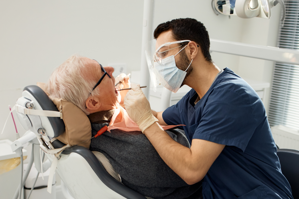
Abfraction is usually diagnosed during a routine dental examination. Dentists will look for visual clues and then confirm the diagnosis with tests. The process of diagnosing abfraction is:
Visual and physical examination
During the visual and physical examination, the dentist will look for these signs:
- Wedge shaped lesions: The dentist will look for the characteristic wedge or V shaped notches at the gumline. Usually these are visible on premolars or canines. The lesions are typically hard and smooth and located at the cervical area where the tooth and gum meet.
- Teeth sensitivity: To assess teeth sensitivity the dentist may apply cold air or light pressure to the area to check for dentin response. When abfraction occurs, the dentin is usually tender and sensitive to touch or temperature.
- Tooth discolouration: If teeth are discoloured at the cervical area it is typically a sign of abfraction. This discolouration is a common symptom caused by exposure of the darker, dentin layer the dentist would notice.
- Tooth fragility: The dentist will note any tooth fragility which may indicate an advanced stage of abfraction with the possibility of tooth loss.
Patient history assessment
- Habits and previous dental and health issues: The dentist will look into the patient’s history both medical and dental. This is an important part of the diagnosis to determine if the patient has a history of bruxism or other factors that may contribute to abfraction issues.
Further tests
- X-rays: The dentist may undertake further tests like x-rays to assess the extent of the damage beneath the gum line. These tests evaluate the health of the jawbone, and rule out other conditions like gum disease.
Abfraction Treatment
Abfraction treatment involves addressing the cause and fixing the damage from the condition. Abfraction cannot be reversed as tooth enamel and dentin are unable to be restored. However, it can be effectively managed and controlled. With treatment and addressing underlying causes, the progression of abfraction can be stopped or slowed down, symptoms relieved and tooth damage repaired. Not all abfraction lesions require immediate treatment but management is important to prevent further tooth damage. After diagnosing abfraction, the dental professional will tailor treatment and management to each patient, depending on the severity and underlying cause. Abfraction treatment includes:
- Monitoring early symptoms: In the initial stages when abfraction lesions are asymptomatic, the dentist may decide to monitor rather than begin treatment. This will occur if the lesion is small, stable, not sensitive and tooth strength is uncompromised. Regular dental checkups will be scheduled to monitor changes in size, depth or symptoms. Preventative advice will also be provided to stop any progression.
- Managing tooth sensitivity: As sensitivity is one of the first symptoms of abfraction, the dentist may manage it by applying fluoride varnish to strengthen the enamel surface or recommend desensitising toothpaste for daily use.
- Bite adjustment: As uneven or excessive force on the teeth is a major cause of abfraction, bite adjustment is an important step in managing the condition. The dentist carefully assesses how the upper and lower teeth contact each other and make adjustments so that force can be distributed more evenly. Usually this is a simple, in-chair procedure which involves reshaping the tooth enamel to create a more streamlined bite. The goal is to remove any interferences and create small, flat areas for the opposing teeth to contact evenly.
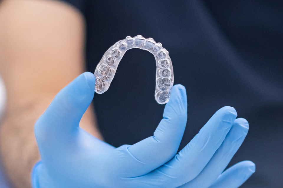
- Mouth guards: For those suffering from bruxism, mouthguards at night help prevent further grinding and clenching. These custom fitted devices protect the teeth from damage caused by the pressure associated with bruxism.
- Orthodontics: In cases where bit misalignment is more extreme, orthodontic treatment may be recommended. Braces and aligners adjust the teeth to evenly distribute the chewing force, stopping the uneven pressure that causes abfraction.
- Dental bonds or fillings: For deep lesions that cause ongoing discomfort, restoration improves the damaged area and prevents further tooth flexure. To restore the area, the dentist will use dental bonds or fillings. Common restorative materials include composite resin, glass ionomer cement or resin modified glass ionomer which rebuild the tooth structure and reduce sensitivity.
- Crowns: In severe cases where a large portion of the tooth structure is lost, a dental crown may be recommended. A crown can treat the damage caused by abfraction by reinforcing the whole tooth and providing strength against biting forces. Crowns will be suggested when 50% of the tooth structure is compromised.
- Veneers: If abfraction affects front teeth, veneers may be an option. A dentist can apply veneers to improve the appearance of the tooth surface but also offer protection against further degradation. Typically veneers are used when aesthetics are the major concern.
- Gum grafting: If gum recession is a contributing factor to abfraction, gum grafting may be a solution. This process involves taking tissue from another area in the mouth and attaching it to the affected site. Gum grafting can help protect the tooth from further damage.
Abfraction Left Untreated
Left untreated, abfraction can lead to permanent serious dental problems. Damage to the tooth can gradually progress over time as abfraction is caused by ongoing mechanical stress. Without intervention and treatment, abfraction can lead to these issues:
- Increased sensitivity: As abfraction progresses, tooth sensitivity tends to increase. As the lesion deepens, the more protective layer of dentin is exposed which leads to more frequent tooth sensitivity. It may begin as mild discomfort but develop into sharp and lingering pain if left unmanaged.
- Tooth structure loss: Without intervention, the enamel at the neck of the tooth becomes thinner and exposes the dentin underneath. Once exposed, tooth structure loss can accelerate.
- Tooth fracture: Left untreated, abfraction can lead to a higher risk of tooth fracture. In severe cases, this can compromise the long term viability of the tooth.
- Gum irritation and recession: As the abfraction lesion enlarges, it can affect the surrounding gum tissue. Food and plaque is more easily trapped in the affected area which can escalate to gum inflammation and eventual recession.
- Tooth loss: In the most extreme cases, tooth loss is a possibility if abfraction continues to remain untreated. While abfraction does not directly cause tooth loss, it does significantly weaken the tooth and increase the likelihood of losing a tooth over time.
- Aesthetic concerns: Abfraction lesions make the tooth look discoloured and often appear as visible notches near the gumline. When these lesions appear on the front teeth, it can significantly affect the appearance of the smile.
How to Prevent Abfraction
Preventing abfraction focuses on reducing excessive tooth pressure, maintaining excellent oral hygiene and regular dental checkups. Strategies to help prevent abfraction include:
- Manage bruxism: For those with tooth clenching or grinding issues, manage bruxism by wearing a night guard at home. Contact the dentist for assessment if you experience jaw pain or headaches as it can be associated with issues like TMJ disorder that is closely linked to abfraction. Botox is another alternative treatment to reduce bruxism.
- Correct bite issues: Have the dentist regularly assess your bite to provide minor adjustments before abfraction develops.
- Use proper brushing techniques: Use a soft bristled toothbrush and brush teeth gently in circular motions. Choose toothpaste that is non-abrasive.
- Strengthen tooth enamel: Consider using fluoride toothpaste or have professional fluoride treatments at the dentist. Avoid excessive teeth whitening procedures which can significantly impact tooth enamel.
- Avoid acidic foods: Limit the amount of food and drink with acids. Rinse your mouth well after acidic intake.
Abfraction and Gum Recession
Abfraction and gum recession are confused because they both affect the gumline, can cause tooth sensitivity and visible changes. Despite the similarities, they are not the same thing. Abfraction is tooth surface loss caused by stress and pressure. It appears as V-shaped notches and tooth discolouration. Gum recession is when the gums pull back exposing the tooth root surface and is primarily caused by advanced gum disease or periodontitis. It is a serious dental issue that involves loss of gum tissue and root exposure but does not directly remove enamel or dentin.
Abfraction is stress-related tooth wear that occurs at the neck of the tooth along the gumline. Although it may resemble gum recession, it is a distinct condition that occurs due to excessive or uneven biting forces. Understanding abfraction is important as it does not resolve on its own and needs to be addressed and managed once symptoms appear. The team at Hawthorn Road Family Dental Clinic are the experts in early diagnosis and treatment of abfraction.